EM Exeter
  • Home
    • Contact us
  • Shop floor
    • Directory
    • Useful links
    • Ortho guidelines
  • Education
    • Diary
    • Doctors >
      • Junior Doctors >
        • Weekly teaching
        • SHOP FLOOR LEARNING
      • Middle grades >
        • Weekly Teaching
        • Procedural sedation
        • Management and leadership
        • MEM:EM Weekly
        • MEM:EM Contents
      • Ultrasound >
        • Ultrasound training
        • VASC Accs/FICB
        • EVE sign up
    • Nurses >
      • Nursing risk assessments
      • Practical Skills resources
      • Training resources
    • Journal club
    • Friday work-out
    • Lunchtime Learning
    • Ophthalmology
    • Quality improvement
    • FOAMed
    • Safeguarding
  • Induction
    • RCEM induction
  • ACADEME
  • Other projects and Social information
    • Social events
    • GreenRDED
    • Comfort Exeter
    • Department newsletter

Lunchtime Learning

LLB - Syncope / presyncope - think WOBBLER

9/17/2020

1 Comment

 
Syncope is a very common presentation to the ED.

The ECG is a vital investigation in the context of a transient loss of consciousness / syncope as it can identify the cause, including rare conditions that can lead to sudden cardiac death.

The pneumonic WOBBLER can be used as a useful way of asessing the ECG in this context with consideration for these rare causes.

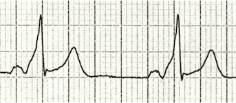
W - Wolff-Parkinson-White syndrome –  A short PR interval and delta wave:
Picture
O - Obstructed AV pathway – look for 2nd or 3rd degree block:
​
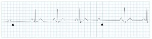
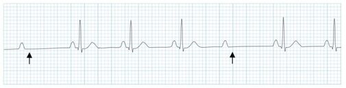
2nd degree block:
Mobitz type 1 - PR prolongs then QRS not present:
Picture
Mobitz 2 - PR Is constant but QRS randomly not present:
Picture
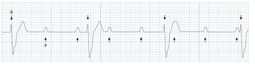
3rd Degree block - complete dissociation between P waves and QRS complexes:
Picture
B - Bifascicular block - The combination of axis deviation and right bundle branch block:
Bifasicular block has a low risk of deteriorating into complete heart block (~ 1% per year) but trifasicular block (Axis deviation, RBBB and prolonged PR) has a greater risk.


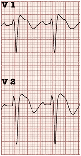
B - Brugada - Coved ST elevation (shark fin) followed by a negative T wave in V1-V3
Picture
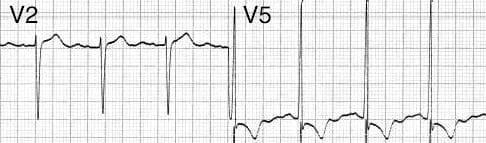
L – Left ventricular hypertrophy - Consider Aortic stenosis and Hypertrophic Obstructive cardiomyopathy (HOCM), these can present with exertional syncope.
​
Picture
LVH diagnostic criteria - S wave in V2 + R wave in V5 > 35 mm
​
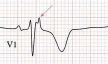
E – Epsilon wave - A feature of Arrythmogenic right ventricular dysplasia (ARVD). This is another rare cause of sudden cardiac arrest.

The epsilon wave looks a bit like the J wave of hypothermia and may be associated with T wave inversion in V1-V3:
Picture
R - Repolarisation abnormality, such as long or Short QT syndrome.
      A prolonged QT is > 440ms in men or > 460ms in women.
      A QT >500ms is associated with increased risk of Torsades de pointes.
      A QT of <350ms is abnormally short.
 

So thats it, next time you see a patient with a transient loss of consiousness make sure thier ECG is not a WOBBLER.

Dr Martin Dore
​
1 Comment
best substance abuse treatment link
4/15/2026 10:44:40 pm

The best substance abuse treatment combines medical detox, therapy, and aftercare support to ensure long-term recovery and prevent relapse.

Reply


Leave a Reply.

    This will be an archive of learning blogs and lunchtime learnings bites!

    Archives

    February 2023
    February 2022
    January 2022
    December 2021
    May 2021
    April 2021
    March 2021
    December 2020
    November 2020
    October 2020
    September 2020
    August 2020
    July 2020
    August 2019

    Categories

    All
    Analgesia
    Cardiovascular
    COVID
    Data Interpretation
    GI
    GI / Surgery
    HEENT Ophthal/ENT/MaxFax
    ID/Immunology/Derm
    Mental Health
    MSK/Ortho/Rheum
    Neuro/Neurosurgery
    Orthopaedics
    Procedures
    Resus
    Resusitation
    Sedation
    Surgery
    Tips And Tricks
    Toxicology/Pharm/Exposure
    Wound Care

    RSS Feed

Proudly powered by Weebly
  • Home
    • Contact us
  • Shop floor
    • Directory
    • Useful links
    • Ortho guidelines
  • Education
    • Diary
    • Doctors >
      • Junior Doctors >
        • Weekly teaching
        • SHOP FLOOR LEARNING
      • Middle grades >
        • Weekly Teaching
        • Procedural sedation
        • Management and leadership
        • MEM:EM Weekly
        • MEM:EM Contents
      • Ultrasound >
        • Ultrasound training
        • VASC Accs/FICB
        • EVE sign up
    • Nurses >
      • Nursing risk assessments
      • Practical Skills resources
      • Training resources
    • Journal club
    • Friday work-out
    • Lunchtime Learning
    • Ophthalmology
    • Quality improvement
    • FOAMed
    • Safeguarding
  • Induction
    • RCEM induction
  • ACADEME
  • Other projects and Social information
    • Social events
    • GreenRDED
    • Comfort Exeter
    • Department newsletter